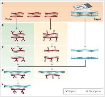
FISH or Fluorescence In Situ Hybridization is a technology that can be employed to visualize specific regions within chromosomes. To accomplish this, a labeled probe and target DNA sequence are required. The labeled probe will either be directly or indirectly labeled with a fluorophore and later detected using fluorescence microscopy.
The first in situ hybridization experiments were done in 1969 by Gall and Pardue. In this initial experiment, a radiolabeled DNA probe hybridized to the DNA of the toad Xenopus, revealing the labeling pattern of DNA that encodes ribosomal RNA. Within their experiment they used both RNA and DNA probes. Fluorescent labels first replaced radioactive labels in 1977 by Rudkin & Stollar. Over the years a variety of modifications to the procedure have given us the current FISH protocol. Some of the defining research in the modern procedure include work by Trask in 2002; and Speicher & Carter in 2005.
In order to use this technique a target region on a chromosome must be selected as well as a probe. The probe is a region of DNA complementary to the region of interest. The probe is then labeled indirectly or directly. To indirectly label a probe, the researcher will label the probe with modified nucleotides that are linked to a hapten (a small molecule that can generate an immune response when bound to a larger molecule). In direct labeling the probe is labeled with nucleotides that are directly linked to a fluorophore. Both the probe and the target are denatured to the point of DNA strand separation. Then the probe will bind to the target DNA sequence. If indirect labeling techniques were previously used, additional reactions are necessary (an enzymatic or immunological detection system is needed.) If the probe was directly labeled, the probe can be detected using a fluorescent microscope.
Indirect Labeling vs. Direct Labeling of the Probe[]
There are advantages and disadvantages to both systems. Indirect labeling requires more time and technical work, but has the potential to generate a brighter signal. The use of other reagents (such as other antibodies) to generate a fluorescence response may reduce the appearance of any background fluorescence. Conversely, direct labeling is fast and easy, but background fluorescence may effect the intensity of the signal.
Other Considerations[]
Include the limitations of the fluorescence microscope - will the researcher be able to see the chromosomes (is the machine sensitive enought?), will the researcher be able to resolve the chromosomes? (are signals that are close together distinct?)
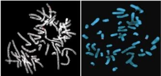
DNA form (is the DNA in mitosis/meiosis or interphase? If the DNA is in interphase DNA will not be as condensed so detecting signal may be challenging, but can be done!)
Diagnose Chromosome abnormalities: Enables clinical diagnoses in instances where patients have an insertion, deletion, duplication, or translocation within their DNA.
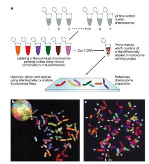
Detect entire chromosomes. (As seen in the figure to the left). Chromosomes will be labeled with a unique color and then hybridized to metaphase chromosomes. The intent of this hybridization is to get an idea of the genetic big picture. So from a clinical standpoint, it displays what chromosomes are present and whether there are any obvious large deletions or translocations. However, this method is not useful for resolving small differences between homologous chromosomes.
Interphase chromosome: useful in detection of chromosomes/gene sequences in solid tumors as these cells do not frequently divide.
Use of FISH in a recent Genetics Publication[]
Repeat Units within the Chinese Watermelon Genome. "The draft genome of watermelon (Citrullus lanatus) and resequencing of 20 diverse accessions" Shaogui Guo et al. Nature Genetics 2012
Nature Genetics recently published a paper on the watermelon genome (November 25, 2012, "The draft genome of watermelon (Citrullus lanatus) and resequencing of 20 diverse accessions"). The genome contains a variety of repeat elements that are found in various locations in the chromosomes. They used FISH to highlight centromere-like repeats (B) telomere-like repeats (C) and ribosomal DNA-like repeats (D).


